 da Re-Fit
Eczema on the Lower Leg That Won't Clear? It May Be Your Veins — Stasis Dermatitis and the Skin Changes of Venous Disease
Itching, red patches, thickening and brown discolouration on the lower leg, repeatedly diagnosed as "eczema" and repeatedly returning after steroid cream. In some such cases the cause may lie not in the skin but in the vein underneath it — and the mechanism is well described.
Recurrent dermatitis on the leg may have a vascular cause
Itching, red patches, thickened skin, brown discolouration around the ankle. Repeated visits to a dermatologist; a diagnosis of "eczema of unknown cause"; steroid cream that works for a while and then does not. If this is familiar, the problem may not be the skin at all but the veins beneath it.
Chronic venous insufficiency (CVI) — of which varicose veins are the commonest expression — is not merely a matter of visible, bulging vessels. When blood refluxes and pools in the leg veins, the skin can eventually be affected, and the result can look very much like a primary skin disease.
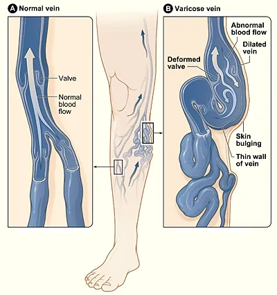
A normal vein (A) and a varicose vein (B). Failed valves allow reflux and the vein wall dilates. (Wikimedia Commons, CC BY-SA)
1. Why look at the veins for a skin problem?
In chronic lower-leg skin changes that do not respond to dermatological treatment, venous reflux under the skin is sometimes the cause. The venous insufficiency produced by varicose veins can lead to a range of skin changes — dermatitis, pigmentation, hardening, and in some cases ulceration.
2. What varicose veins are
Varicose veins develop when the valves inside the leg veins fail. Healthy valves open only toward the heart; when they weaken, blood refluxes downward and pressure inside the vein rises abnormally — venous hypertension.
The condition is very common. In the Vein Consult Program, an international survey of 91,545 adults attending general practitioners, 63.9% had some clinical sign of chronic venous disorder (CEAP C1–C6); the severe stages with skin changes or ulcer (C4–C6) occurred equally in men and women. Meissner and colleagues put varicose veins without skin changes at around 20% of Western populations and active ulcers at up to 0.5%.
3. How venous disease damages the skin — the mechanism
The process is stepwise. Understanding the sequence explains why a vascular problem presents as a dermatological one.
Venous pressure rises → capillaries leak
Reflux raises pressure in the veins, and that pressure is transmitted upstream into the capillaries and venules. Their walls stretch, and plasma, protein and even red blood cells leak into the surrounding skin (Bergan et al., 2008; Silverberg et al., 2023).
Haemosiderin deposition → brown pigmentation
Red cells that escape into the tissue break down and leave behind haemosiderin, an iron-containing pigment. Deposited in the dermis, it produces the characteristic brown or tan discolouration of the lower leg.
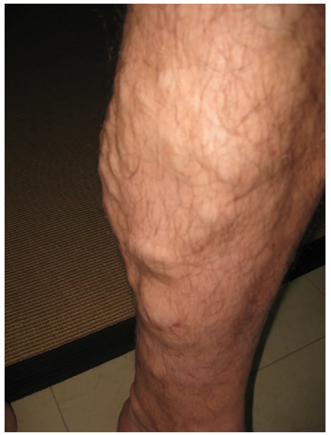
Varicose veins. Valve failure dilates the vein, which becomes tortuous and visible beneath the skin. (Wikimedia Commons / OpenStax, CC BY 4.0)
Leukocyte trapping → chronic inflammation
As flow slows, white blood cells adhere to the microvascular walls. Coleridge Smith and colleagues proposed in the BMJ in 1988 that these trapped white cells occlude capillaries and render the skin ischaemic — the "white cell trapping" hypothesis. The trapped cells activate and release inflammatory cytokines and oxidative stress mediators, sustaining low-grade chronic inflammation (Silverberg et al., 2023; Raffetto et al., 2020).
Fibrin cuffs → impaired tissue oxygenation
Fibrinogen leaking from plasma is deposited around the capillaries as a fibrin cuff — the mechanism Browse and Burnand proposed in the Lancet in 1982. As the cuff thickens it impedes the passage of oxygen and nutrients from capillary to skin, and skin cells are damaged.
Lipodermatosclerosis → fibrosis and hardening
Repeated over years, this inflammation leads to fibrosis of the skin and subcutaneous fat: lipodermatosclerosis. The skin becomes hard and thick, characteristically above the inner ankle. We have described how oedema becomes fibrosis in detail elsewhere.
Venous ulcer → a chronic wound
If the process continues over a long period, a venous leg ulcer can develop, usually around the inner ankle. Venous ulcers are among the most common ulcers of the lower extremity (Raffetto et al., 2020), and they are slow to heal and quick to recur.
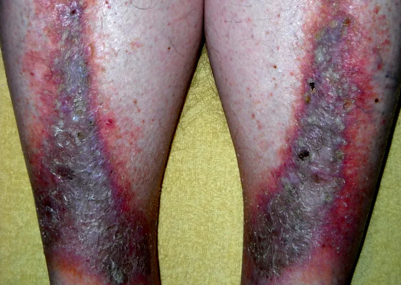
Stasis dermatitis. Venous stasis from varicose veins produces pigmentation and eczematous change. (Wikimedia Commons, CC BY-SA 3.0)
4. The skin changes associated with venous disease
Brown pigmentation (haemosiderin staining) — tan to brown discolouration at the ankle and lower calf.
Eczema / dermatitis (stasis dermatitis) — itching, redness, weeping, dryness and cracking.
Hardening and thickening (lipodermatosclerosis) — firm, bound-down skin above the inner ankle.
White patches (atrophie blanche) — ivory-white scarred areas with loss of the small vessels at their centre.
Chronic wound (venous ulcer) — shallow, broad ulceration around the inner ankle.
Dilated surface vessels (telangiectasia) — red thread veins and spider veins.
5. Ordinary eczema or venous dermatitis — how to tell them apart
Ordinary eczema or contact dermatitis can appear anywhere on the body, is often linked to an identifiable trigger, is rarely accompanied by swelling, and responds well to antihistamines and topical steroids.
Venous (stasis) dermatitis is confined to the ankle and lower leg, worsens with prolonged standing or activity, comes with ankle swelling and heaviness, shows brown pigmentation, and often does not clear with steroids alone.
Stasis dermatitis occurs mainly in adults over 40, only on the lower legs, with brown pigmentation. Atopic dermatitis usually begins in childhood and favours the inner elbows and backs of the knees.
One further point that matters for anyone who has been treating their leg with creams for months. Chronically inflamed skin is more easily sensitised to allergens that intact skin would tolerate (Nedorost & Hammond, 2020). Stasis dermatitis therefore frequently acquires a secondary allergic contact dermatitis to the very products being applied to it — which is one reason "eczema of the lower leg" so often worsens despite treatment.
Consider a consultation with a vascular surgeon or a cardiovascular and thoracic surgeon if you have: eczema or itching that recurs only on the ankle and lower calf · leg swelling that worsens in the evening · brown discolouration around the ankle lasting months · skin that feels hard or bound down · a wound near the ankle that will not heal · visible tortuous veins on the leg.
6. Diagnosis of venous dermatitis
The core investigation is duplex ultrasound, which establishes whether reflux is present and for how long, whether the saphenous valves are competent, and whether there is deep vein thrombosis. It is non-invasive and involves no radiation. The Society for Vascular Surgery / American Venous Forum guideline recommends it for every patient with varicose veins or more advanced venous disease (GRADE 1A).
If duplex ultrasound shows reflux or valve incompetence, stasis dermatitis can develop even when no varicose veins are visible on the surface.
7. Treatment: looking at the vein as well as the skin
Whether skin changes are due to venous insufficiency has to be confirmed by examination; where they are, treatment of the underlying reflux is considered as well.
Compression therapy. Medical compression stockings reduce venous pressure. This is the basic conservative treatment, and for venous ulcers the guideline recommends it as the primary treatment to aid healing (GRADE 1B).
Sclerotherapy. A sclerosant injected into refluxing veins closes them; used mainly for small and medium-sized varicosities.
Endovenous thermal ablation. Laser or radiofrequency closes the refluxing vein from inside through a needle puncture rather than an open incision.
Surgery (high ligation and stripping). The origin of reflux is tied off and the dilated vein removed; reserved for selected cases.
The strongest guideline statement in this area concerns ulcers: to reduce recurrence of venous ulcers, ablation of the incompetent superficial veins is recommended in addition to compression (GRADE 1A). The recommendation, in other words, is to treat the vein as well as the wound.
Where venous reflux is the cause, the skin changes can be expected to improve gradually over months once the reflux has been treated. The degree of improvement varies from person to person.
Key points: varicose veins and skin changes
Recurrent skin changes on the lower leg — pigmentation around the ankle, eczema that will not clear — warrant a check of the veins. Varicose veins are not only a cosmetic issue; they are a condition that can damage the skin and the tissue beneath it.
If a chronic leg skin problem is not resolving with dermatological treatment, it is worth having the venous function assessed with a duplex ultrasound.
References
- Rabe E, Guex JJ, Puskas A, et al. Epidemiology of chronic venous disorders in geographically diverse populations: results from the Vein Consult Program. Int Angiol. 2012;31(2):105-15. (PMID: 22466974)
- Meissner MH, Gloviczki P, Bergan J, et al. Primary chronic venous disorders. J Vasc Surg. 2007;46 Suppl S:54S-67S. (PMID: 18068562)
- Bergan JJ, Pascarella L, Schmid-Schönbein GW. Pathogenesis of primary chronic venous disease: Insights from animal models of venous hypertension. J Vasc Surg. 2008;47(1):183-92. (PMID: 18178472)
- Silverberg J, Jackson JM, Kirsner RS, et al. Narrative Review of the Pathogenesis of Stasis Dermatitis: An Inflammatory Skin Manifestation of Venous Hypertension. Dermatol Ther (Heidelb). 2023;13(4):935-950. (PMID: 36949275)
- Raffetto JD, Ligi D, Maniscalco R, Khalil RA, Mannello F. Why Venous Leg Ulcers Have Difficulty Healing: Overview on Pathophysiology, Clinical Consequences, and Treatment. J Clin Med. 2020;10(1):29. (PMID: 33374372)
- Browse NL, Burnand KG. The cause of venous ulceration. Lancet. 1982;2(8292):243-5. (PMID: 6124673)
- Coleridge Smith PD, Thomas P, Scurr JH, Dormandy JA. Causes of venous ulceration: a new hypothesis. Br Med J (Clin Res Ed). 1988;296(6638):1726-7. (PMID: 3135894)
- Nedorost S, Hammond M. Art of prevention: Allergic sensitization through damaged skin: Atopic, occupational, and stasis dermatitis. Int J Womens Dermatol. 2020;6(5):381-383. (PMID: 33898703)
- Gloviczki P, Comerota AJ, Dalsing MC, et al. The care of patients with varicose veins and associated chronic venous diseases: clinical practice guidelines of the Society for Vascular Surgery and the American Venous Forum. J Vasc Surg. 2011;53(5 Suppl):2S-48S. (PMID: 21536172)
Frequently asked questions
- Can stasis dermatitis occur without visible varicose veins?
- If duplex ultrasound shows reflux or valve incompetence, stasis dermatitis can develop even when no varicose veins are visible on the surface.
- How can ordinary eczema be told apart from venous (stasis) dermatitis on the lower leg?
- Venous (stasis) dermatitis is confined to the ankle and lower leg, worsens with prolonged standing or activity, comes with ankle swelling and heaviness, shows brown pigmentation, and often does not clear with steroids alone.
- How is stasis dermatitis distinguished from atopic dermatitis?
- Stasis dermatitis occurs mainly in adults over 40, only on the lower legs, with brown pigmentation. Atopic dermatitis usually begins in childhood and favours the inner elbows and backs of the knees.
- Why does the skin around the ankle turn brown with varicose veins?
- Red cells that escape into the tissue break down and leave behind haemosiderin, an iron-containing pigment. Deposited in the dermis, it produces the characteristic brown or tan discolouration of the lower leg.
- How is venous disease diagnosed when the lower-leg skin changes?
- The core investigation is duplex ultrasound, which establishes whether reflux is present and for how long, whether the saphenous valves are competent, and whether there is deep vein thrombosis. It is non-invasive and involves no radiation.

Don't navigate your leg symptoms alone
From precise duplex ultrasound diagnosis to treatment — consult directly with board-certified surgeons trained at Asan Medical Center.
Language support is available through our interpretation system.