 da Re-Fit
Why Are Leg Veins Examined With Doppler Ultrasound? Can't CT or MRI Do It?
When you come in for a varicose vein evaluation, the first test is ultrasound, not CT or MRI. Venous reflux is confirmed by watching the direction of blood flow while you stand. This article explains what international guidelines recommend, what CT and MR venography can and cannot show, and when CT or MRI is actually needed.
"Shouldn't I have a CT or MRI for a more detailed look?"
"Is it all right to decide on a procedure with only an ultrasound?"
These are common questions in a vein consultation. For many other conditions CT or MRI is the more detailed test, so it is natural to assume the same applies to leg veins.
For leg veins the order is different. Doppler ultrasound is the basic test, and CT or MRI is added in specific situations. The reason is not convenience. The information needed to diagnose varicose veins is the information ultrasound provides.
1. What matters is the direction of blood flow, more than the shape of the vein
Leg veins contain valves that let blood travel only upward, toward the heart. Varicose veins and chronic venous insufficiency begin when those valves do not close properly and blood flows back toward the foot. This is called reflux.
The central question in diagnosing varicose veins is whether the valves are working, in other words whether blood is flowing backward. Whether a vein is enlarged and where it runs also matter, but they do not complete the diagnosis.
The test commonly called "Doppler ultrasound" is formally duplex ultrasound. It combines an ordinary ultrasound image, which shows the vein's cross-section and course, with a Doppler function that detects the movement of blood. Doppler ultrasound shows the direction and duration of blood flow in real time. It can measure directly whether reflux is present, in which vein and segment, and for how long.
2. Reflux appears when you stand, and it has to be provoked
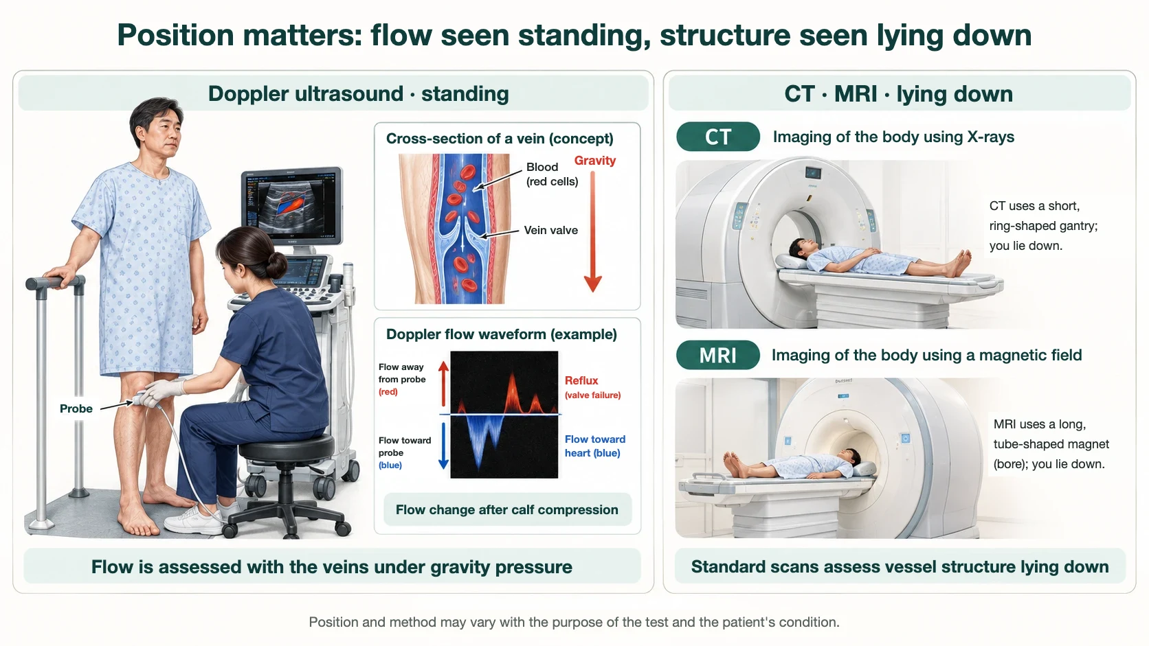
Reflux is not readily seen when you lie still. When you stand, gravity puts pressure on the leg veins, and if the valves cannot hold, blood flows downward. During the ultrasound examination you stand while the examiner squeezes and releases your calf, or asks you to bear down, and the flow at that moment is measured.
A 2003 study that established the reflux criteria examined the same veins in the standing and supine positions. Among veins in healthy volunteers, 48 segments showed reflux longer than 0.5 seconds while standing, and 6 of them (13%) measured under 0.5 seconds when supine. In the other direction, of 37 segments over 0.5 seconds when supine, 22 (59%) were under 0.5 seconds when standing. Position can change the result in both directions, and the authors concluded that reflux testing should be performed with the patient standing (Labropoulos et al., 2003).
The guidelines say the same. The European guideline describes reflux assessment as preferably done upright, with reflux provoked. The American guideline advises examining the patient standing whenever possible, and allows a sitting or reverse Trendelenburg position for patients who cannot stand.
CT and MRI are acquired while you lie still on a table. They record the shape of the veins at one moment, in a position where the veins are not under pressure. Provoking reflux and measuring that flow is not part of a standard CT or MRI scan.
The reflux cut-off values are explained in our article on the 0.5-second reflux standard.
3. International guidelines recommend ultrasound as the basic test
Several society guidelines agree on this point.
- The European Society for Vascular Surgery (ESVS) 2022 guidelines recommend full lower limb duplex ultrasound as the primary imaging modality for diagnosis and treatment planning in patients with suspected or clinically evident chronic venous disease (Class I, Level B).
- The Society for Vascular Surgery, American Venous Forum, and American Vein and Lymphatic Society (SVS/AVF/AVLS) guidelines recommend duplex ultrasound as the diagnostic test of choice to evaluate for venous reflux in patients with chronic venous disease of the lower extremities (strong recommendation, Level B evidence).
- The American College of Radiology (ACR) Appropriateness Criteria rate lower extremity duplex ultrasound as "usually appropriate" for the initial diagnosis of varicose veins, and CT venography and MR venography of the lower extremity as "usually not appropriate."
The third document comes from the society of the specialists who perform and read CT and MRI. Radiologists, too, name ultrasound as the first test for varicose veins.
4. What CT shows in varicose veins, and what it does not
CT venography can image leg veins. In a study of 100 patients with varicose veins at a Korean university hospital, three-dimensional CT venography visualized the entire length of the great saphenous vein in 99.5% of 200 veins. Showing the course and connections of the veins in a single three-dimensional image is a strength of CT (Lee et al., 2008).
What CT shows, however, is the shape and size of the vein. In the same study, great saphenous veins with reflux on ultrasound had a mean diameter of 7.0 mm on CT, and those without reflux 4.9 mm. Predicting reflux from diameter gave a sensitivity of 98.2% and a specificity of 83.3%. Another Korean study, using a great saphenous vein diameter of about 5.9 mm as the cut-off, reported a sensitivity of 91.4% and a specificity of 81.8% (Choi et al., 2022).
These results can be read two ways. Enlarged veins often have reflux, and diameter alone misclassifies some veins. Some veins are large without reflux. In both studies the reference test that determined reflux was ultrasound. CT estimates reflux from size. Ultrasound measures it.
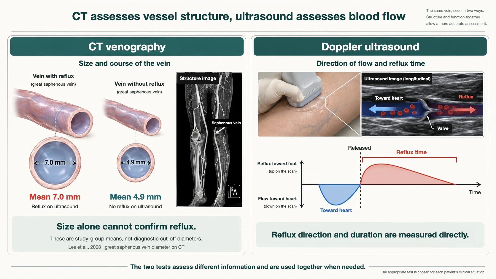
A Japanese study that used CT venography for preoperative assessment describes the same division of roles: CT for anatomy, with ultrasound focused on hemodynamic assessment (Sato et al., 2011). The two tests do not replace each other. They look at different things.
There is also a difference in burden. The European guideline states that CT venography requires iodinated contrast and ionizing radiation. Ultrasound uses neither, so it can be repeated before treatment, during the procedure, and at follow-up.
| Doppler (duplex) ultrasound | CT venography | MR venography | |
|---|---|---|---|
| Position | Can be done standing | Supine | Supine |
| Reflux | Direction and duration measured directly | Estimated from size and shape | Limited with standard imaging |
| Radiation | None | Yes | None |
| Contrast | Not used | Iodinated contrast | Depends on the technique |
| Best seen | Superficial and deep veins of the leg | Pelvic and abdominal veins, overall course | Pelvic and abdominal veins, overall course |
5. There are cases where CT or MRI is needed
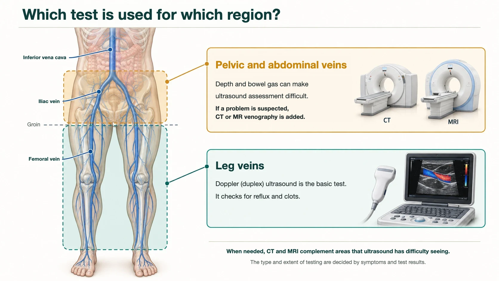
There are situations where ultrasound alone is not enough. These involve the veins above the groin, in the pelvis and abdomen.
Pelvic and abdominal veins lie deep and can be hidden by bowel gas. The European guideline notes that abdominal ultrasound can be technically difficult in patients with abdominal obesity or bowel gas, and that ultrasound of the pelvic veins requires specific expertise.
The same guideline advises suspecting a problem above the groin when there is extensive swelling of one whole leg, a venous ulcer, or collateral veins on the abdominal wall, or when leg ultrasound shows an abnormal flow pattern in the femoral vein. When an intervention is being considered in such patients, it recommends cross-sectional imaging by MR venography or CT in addition to ultrasound (Class I, Level C).
The ACR criteria likewise rate CT venography and MR venography of the abdomen and pelvis as "usually appropriate" when leg varicose veins of suspected pelvic origin, or iliac or inferior vena cava disease, are in question. Leg ultrasound remains part of the evaluation in these cases as well.
In short, CT and MRI do not replace ultrasound. They complement it where ultrasound has difficulty reaching.
6. For leg clots, ultrasound also comes first
Alongside reflux, a vein examination checks for deep vein thrombosis. The examiner presses on the vein with the probe and looks at whether it compresses completely.
In a meta-analysis of 100 cohorts, with contrast venography as the reference, ultrasound had a sensitivity of 94.2% and a specificity of 93.8% for proximal deep vein thrombosis above the knee. For distal clots in the calf, sensitivity was lower at 63.5% (Goodacre et al., 2005).
A meta-analysis of MR venography reported a sensitivity of 93% and a specificity of 96%. The figures are high, but the studies varied widely, and the authors concluded that the evidence did not support replacing ultrasound with MR venography as the first-line test. They added that it may be an alternative for patients in whom ultrasound is inadequate or not feasible (Abdalla et al., 2015). The order is the same for clots: ultrasound first, with CT or MRI added when needed.
The relationship between varicose veins and clots is covered in our article on varicose veins and deep vein thrombosis.
7. Ultrasound has limits too
Ultrasound does not show everything.
- Results can vary with the examiner. CT and MRI are acquired with a set protocol, whereas ultrasound is a real-time search with a hand-held probe. This is one reason the International Union of Phlebology (UIP) issued a separate consensus document on how to perform, interpret and report the examination and how to train examiners.
- Pelvic and abdominal veins can be hard to see. As described above, cross-sectional imaging may be needed here.
- Small calf clots can be missed. In the meta-analysis above, sensitivity for distal clots was lower than for proximal ones.
- Finding reflux is not the same as finding the cause of symptoms. Even when ultrasound confirms reflux, the location and pattern of symptoms and the physical examination have to be considered together.
So in an ultrasound examination, the position, the extent of the scan and the method used matter as much as the equipment. The extent of the scan and the process of building a vein map are explained in our article on vein mapping.
8. In summary
- In varicose veins the question is whether blood flows backward, and this is measured by provoking reflux while you stand.
- Doppler ultrasound measures the direction and duration of that reflux directly, and because it uses no radiation or contrast it can be repeated.
- CT and MR venography show the overall course of the veins and the pelvic and abdominal veins, but because they are acquired lying down they are not used to measure reflux directly.
- If obstruction of the veins above the groin or a pelvic vein problem is suspected, CT or MRI may be needed in addition to ultrasound.
You may want to ask these questions at your examination.
- "Is the reflux test done standing?"
- "In which vein was reflux found, and for how many seconds?"
- "Were the deep veins and possible clots checked as well?"
- "Is there any finding in my case that calls for an additional test such as CT or MRI?"
At da Re-Fit Clinic we consider the ultrasound findings together with your symptoms when deciding on treatment, and when a finding is difficult to judge by ultrasound alone, we explain why an additional test is needed.
References
- De Maeseneer MG, Kakkos SK, Aherne T, et al. European Society for Vascular Surgery (ESVS) 2022 Clinical Practice Guidelines on the Management of Chronic Venous Disease of the Lower Limbs. Eur J Vasc Endovasc Surg. 2022;63(2):184-267. (PMID: 35027279) — duplex ultrasound recommended as the primary imaging modality (Recommendation 3); cross-sectional imaging for suspected supra-inguinal obstruction (Recommendation 5).
- Gloviczki P, Lawrence PF, Wasan SM, et al. The 2022 Society for Vascular Surgery, American Venous Forum, and American Vein and Lymphatic Society clinical practice guidelines for the management of varicose veins of the lower extremities. Part I. Duplex Scanning and Treatment of Superficial Truncal Reflux. J Vasc Surg Venous Lymphat Disord. 2023;11(2):231-261. (PMID: 36326210) — duplex ultrasound recommended to evaluate venous reflux; examination in the standing position.
- Rochon PJ, Reghunathan A, Kapoor BS, et al. ACR Appropriateness Criteria® Lower Extremity Chronic Venous Disease. J Am Coll Radiol. 2023;20(11S):S481-S500. (PMID: 38040466) — appropriateness ratings of imaging by clinical scenario.
- Labropoulos N, Tiongson J, Pryor L, et al. Definition of venous reflux in lower-extremity veins. J Vasc Surg. 2003;38(4):793-798. (PMID: 14560232) — reflux cut-off values by vein; standing versus supine comparison.
- Coleridge-Smith P, Labropoulos N, Partsch H, Myers K, Nicolaides A, Cavezzi A. Duplex ultrasound investigation of the veins in chronic venous disease of the lower limbs — UIP consensus document. Part I. Basic principles. Vasa. 2007;36(1):53-61. (PMID: 17323300) — International Union of Phlebology consensus on examination method, interpretation, reporting and training.
- Lee W, Chung JW, Yin YH, et al. Three-Dimensional CT venography of varicose veins of the lower extremity: image quality and comparison with doppler sonography. AJR Am J Roentgenol. 2008;191(4):1186-1191. (PMID: 18806163) — image quality of 3D CT venography and comparison with ultrasound findings.
- Choi JY, Lee JH, Kwon OJ. Association between the saphenous vein diameter and venous reflux on computed tomography venography in patients with varicose veins. PLoS One. 2022;17(2):e0263513. (PMID: 35167584) — saphenous vein diameter on CT and reflux on ultrasound.
- Sato K, Orihashi K, Takahashi S, et al. Three-dimensional CT Venography: A Diagnostic Modality for the Preoperative Assessment of Patients with Varicose Veins. Ann Vasc Dis. 2011;4(3):229-234. (PMID: 23555458) — roles of CT venography and ultrasound in preoperative assessment.
- Goodacre S, Sampson F, Thomas S, van Beek E, Sutton A. Systematic review and meta-analysis of the diagnostic accuracy of ultrasonography for deep vein thrombosis. BMC Med Imaging. 2005;5:6. (PMID: 16202135) — diagnostic accuracy of ultrasound for deep vein thrombosis.
- Abdalla G, Fawzi Matuk R, Venugopal V, et al. The diagnostic accuracy of magnetic resonance venography in the detection of deep venous thrombosis: a systematic review and meta-analysis. Clin Radiol. 2015;70(8):858-871. (PMID: 26091753) — diagnostic accuracy of MR venography for deep vein thrombosis.
This article provides general medical information. Which tests are needed, and how extensive they should be, depend on your symptoms and veins and need to be confirmed in consultation.
Frequently asked questions
- Is ultrasound enough to diagnose varicose veins, or is CT or MRI needed?
- Doppler ultrasound is the basic test, and CT or MRI is added in specific situations. The reason is not convenience. The information needed to diagnose varicose veins is the information ultrasound provides.
- Why is the Doppler ultrasound for varicose veins done standing?
- Reflux is not readily seen when you lie still. When you stand, gravity puts pressure on the leg veins, and if the valves cannot hold, blood flows downward.
- Can CT or MRI measure venous reflux in varicose veins?
- CT and MRI are acquired while you lie still on a table. They record the shape of the veins at one moment, in a position where the veins are not under pressure. Provoking reflux and measuring that flow is not part of a standard CT or MRI scan.
- When is CT or MRI needed in addition to ultrasound for varicose veins?
- If obstruction of the veins above the groin or a pelvic vein problem is suspected, CT or MRI may be needed in addition to ultrasound.
- What are the limits of Doppler ultrasound for leg veins?
- Results can vary with the examiner. Pelvic and abdominal veins can be hard to see. Small calf clots can be missed.

Don't navigate your leg symptoms alone
From precise duplex ultrasound diagnosis to treatment — consult directly with board-certified surgeons trained at Asan Medical Center.
Language support is available through our interpretation system.