 da Re-Fit
How Does Doppler Ultrasound See Blood Flow? A Plain-Language Explanation for Patients
Have you wondered about the red and blue on the screen during an ultrasound, the whooshing sound from the speaker, or why your calf is squeezed and released? From the Doppler effect explained with an ambulance siren to color Doppler, waveforms and the reason for the gel, this article explains how a venous ultrasound examination works.
Questions come up during an ultrasound examination.
"There are flashes of red and blue on the screen. What are they?"
"The machine makes a whooshing sound. Is that my blood flowing?"
"Why do you keep squeezing and releasing my calf?"
These are hard to ask in the middle of a scan, so this article answers them. Once you know how it works, the screen looks different.
1. Ultrasound is sound you cannot hear
Ultrasound is sound pitched too high for the human ear. We hear sounds that vibrate up to about 20,000 times per second. The ultrasound used in an examination vibrates several million times per second.
It works like an echo. Shout across a valley and the sound bounces back from the other side. The longer the echo takes to return, the farther away it was reflected.
The hand-held part of the ultrasound machine is called the probe. The probe sends ultrasound into the body and receives the echoes that bounce back from the tissues. The machine uses the return time of each echo for depth and its strength for brightness, and draws a black-and-white image. The size and position of a vein, the shape of its wall, and whether there is a clot are seen on this black-and-white image.
The test does not use X-rays, so there is no radiation exposure.
2. The Doppler effect, explained with an ambulance siren
The black-and-white image alone cannot show which way the blood inside a vessel is flowing. That takes the Doppler effect.
An ambulance siren sounds higher as it approaches and lower as it passes and moves away. The siren itself has not changed, but the pitch you hear has. When the source of a sound is approaching, the sound waves are packed closer together, and when it is moving away they are spread apart.
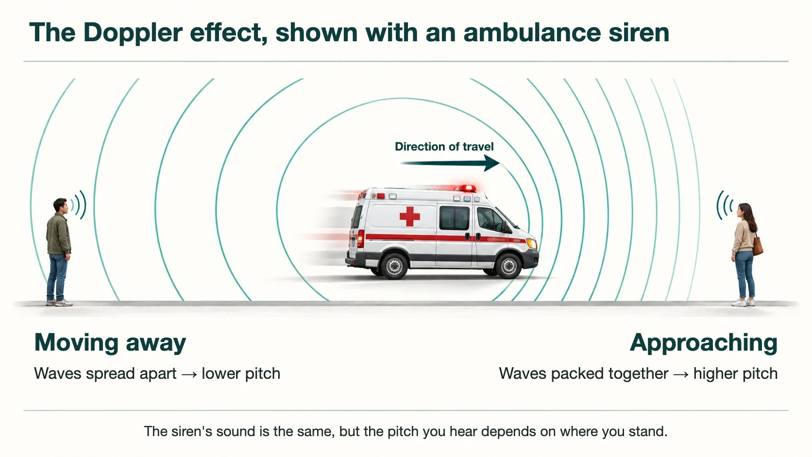
The Austrian physicist Christian Doppler published this principle in 1842, and it carries his name. It was confirmed at the time with trumpets played on a moving train, comparing the pitch as the train approached and moved away (Bollinger and Partsch, 2003).
3. Inside a blood vessel, red blood cells play the part of the siren
Red blood cells are carried along in the blood. The ultrasound sent by the probe bounces back from these cells.
- If the red blood cells are moving toward the probe, the returning ultrasound is higher in pitch than what was sent.
- If they are moving away from the probe, the returning ultrasound is lower in pitch.
- The faster they move, the larger the difference in pitch.
The machine calculates the difference in pitch between the ultrasound it sent and the ultrasound that came back. Doppler ultrasound reads the change in pitch of ultrasound that bounces back from moving red blood cells, and from it works out which way the blood is flowing and how fast.
The first person to apply this principle to the human body was Shigeo Satomura in Japan in the 1950s. He obtained Doppler signals from the movement of the heart using ultrasound, and then, with a colleague, built a device that recorded blood flow in arteries and veins from the surface of the skin (Coman and Popescu, 2015).
4. What are the red and blue on the screen?
A change in pitch shown only as numbers would be hard to follow. So the machine colors the parts where blood is moving and lays that color over the black-and-white image. This is called color Doppler.
Many people assume red means artery and blue means vein, because that is how textbook diagrams are drawn. But red and blue on the screen do not mean artery and vein. They show whether blood is moving toward the probe or away from it.
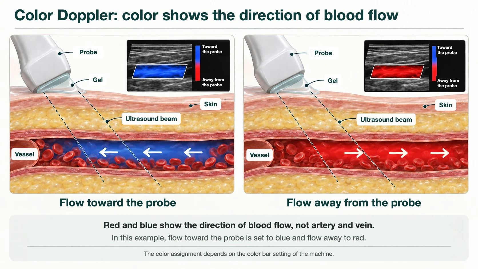
Which color stands for which direction is shown on a color bar at the side of the screen, and the examiner can change it. What matters is not the color itself but the moment the color changes.
In a normal vein, blood flows only toward the heart, so only one color appears. In a vein whose valves do not close properly, blood moves up toward the heart and then comes back down toward the foot. On the screen, a vein that showed one color switches to the opposite color. The direction of flow has reversed within the same vein, and that is reflux.
5. The whooshing sound and the wave-shaped graph
Color Doppler shows at a glance whether reflux is present, but it is not well suited to measuring exactly how long it lasts. For that, the examiner picks one point in the vein and plots the flow there over time.
The graph has a baseline across the middle. Flow in one direction is drawn above the baseline, and flow in the opposite direction below it. The horizontal axis is time.
The whooshing sound you hear comes from here as well. It is not a microphone picking up the sound of your blood. The difference in pitch between the ultrasound sent and the ultrasound returned happens to fall within the range of human hearing, so the machine plays that difference through a speaker. Fast flow gives a higher sound and slow flow a lower one.
6. Why you stand, and why your calf is squeezed and released
When you stand still, blood in the veins moves very slowly. To find out whether the valves close properly, they have to be tested.
When the examiner squeezes your calf, the blood inside is pushed up toward the heart. When the hand is released, gravity pulls that blood back down. A healthy valve closes at once and holds the blood. A valve that is not doing its job lets the blood run down toward the foot.
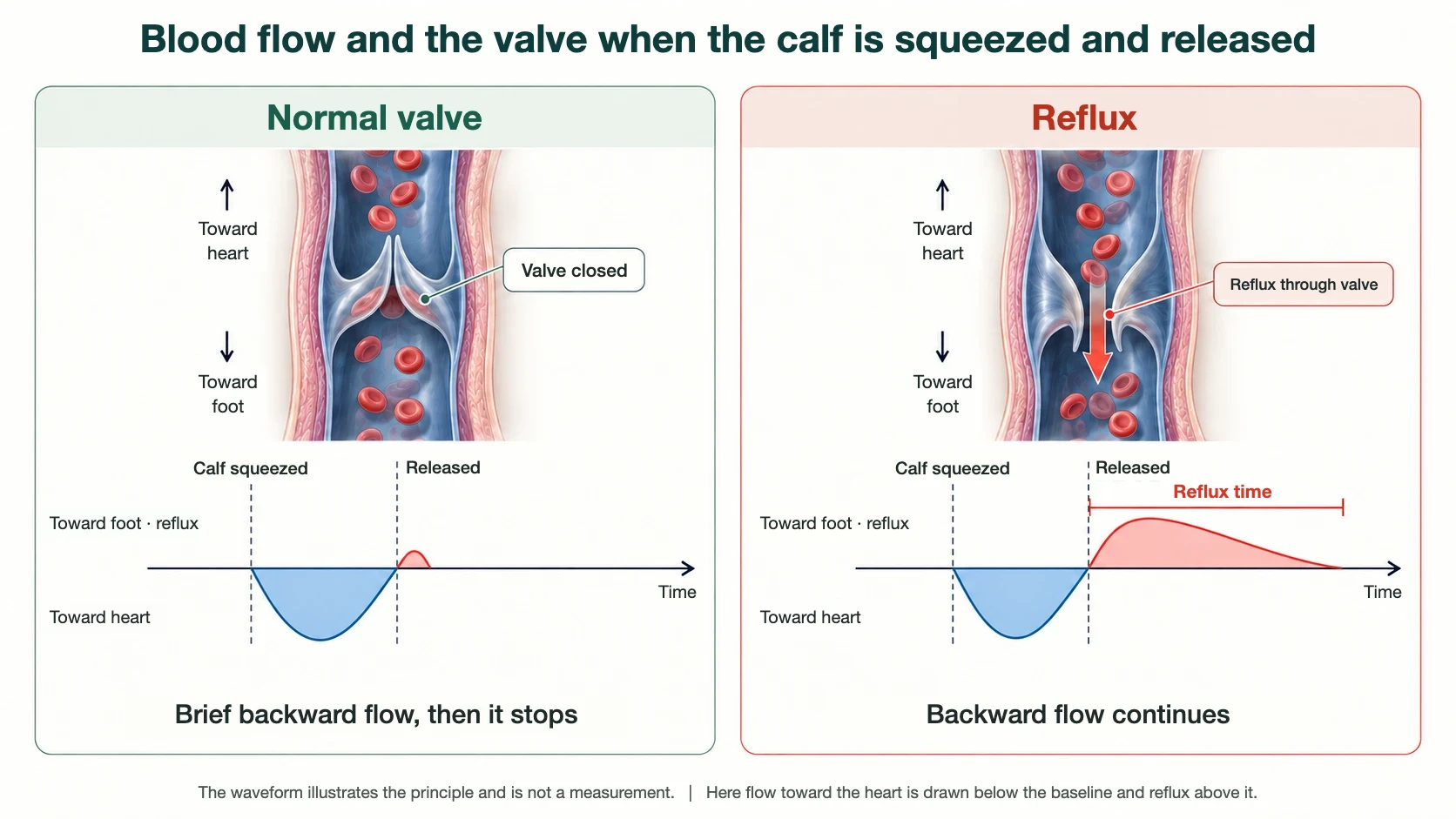
In a vein examination, the waveform is used to measure how long blood keeps flowing backward toward the foot after the calf is squeezed and released. Even a healthy valve takes a very short time to close, so a brief moment of backward flow is normal. For that reason each type of vein has its own cut-off time, and 0.5 seconds is used for the veins near the surface (Labropoulos et al., 2003). How that cut-off was established is explained in our article on the 0.5-second reflux standard.
The reason for standing is the same. Only when you stand does gravity act on the leg veins, so the valves are tested under the conditions they face in daily life. The European Society for Vascular Surgery guideline also describes reflux assessment as preferably done in the upright position.
7. Why gel is applied, and why the probe is tilted
The gel gives the ultrasound a path. Ultrasound does not pass through air well. Even a thin layer of air between the probe and the skin reflects most of it before it enters the body. The gel fills that gap.
Tilting the probe has to do with the nature of the Doppler effect. When an ambulance drives straight toward you the pitch changes clearly, but at the moment it crosses directly in front of you there is almost no change. A blood vessel is the same. If the ultrasound meets the vessel at a right angle, the blood is neither approaching nor moving away from the probe, and no signal is picked up. So the examiner sets the angle of the probe so that the ultrasound meets the flow at a slant.
For the same reason, Doppler ultrasound is a test in which the examiner's hands and judgment affect the result. The waveform can vary with the angle of the probe, the point measured and the way the calf is squeezed. This is why the International Union of Phlebology (UIP) issued a consensus document to standardize how the examination is performed.
8. In summary
- Ultrasound is high-pitched sound you cannot hear, and it draws an image of the inside of the body from echoes.
- Ultrasound that bounces off moving red blood cells comes back at a different pitch. That change gives the direction and speed of the blood.
- Red and blue on the screen show the direction of flow, not artery and vein. When the color reverses within the same vein, reflux is suspected.
- Squeezing and releasing the calf tests the valves, and the duration of backward flow is measured on the waveform.
At your next examination, feel free to watch the screen. If you look at whether the color changes the moment the hand leaves your calf, and how far the graph extends on the other side of the baseline, the explanation of your results will be easier to follow.
Whether CT or MRI can be used instead of ultrasound is covered in Why Are Leg Veins Examined With Doppler Ultrasound?
References
- Bollinger A, Partsch H. Christian Doppler is 200 years young. Vasa. 2003;32(4):225-233. (PMID: 14694774) — publication and confirmation of the Doppler principle and its application to vascular diagnosis.
- Coman IM, Popescu BA. Shigeo Satomura: 60 years of Doppler ultrasound in medicine. Cardiovasc Ultrasound. 2015;13:48. (PMID: 26699126) — the first medical applications of Doppler ultrasound.
- Coleridge-Smith P, Labropoulos N, Partsch H, Myers K, Nicolaides A, Cavezzi A. Duplex ultrasound investigation of the veins in chronic venous disease of the lower limbs — UIP consensus document. Part I. Basic principles. Vasa. 2007;36(1):53-61. (PMID: 17323300) — International Union of Phlebology consensus on how to perform the examination.
- Labropoulos N, Tiongson J, Pryor L, et al. Definition of venous reflux in lower-extremity veins. J Vasc Surg. 2003;38(4):793-798. (PMID: 14560232) — reflux cut-off times by vein.
- De Maeseneer MG, Kakkos SK, Aherne T, et al. European Society for Vascular Surgery (ESVS) 2022 Clinical Practice Guidelines on the Management of Chronic Venous Disease of the Lower Limbs. Eur J Vasc Endovasc Surg. 2022;63(2):184-267. (PMID: 35027279) — patient position and provocation of reflux in duplex ultrasound.
This article provides general medical information. How the examination is performed and how the results are interpreted depend on your condition and need to be confirmed in consultation.
Frequently asked questions
- What do red and blue mean on a Doppler ultrasound of the leg veins?
- Red and blue on the screen show the direction of flow, not artery and vein. When the color reverses within the same vein, reflux is suspected.
- Why is the calf squeezed and released during a Doppler ultrasound for varicose veins?
- Squeezing and releasing the calf tests the valves, and the duration of backward flow is measured on the waveform.
- Why is a Doppler ultrasound for varicose veins done standing up?
- Only when you stand does gravity act on the leg veins, so the valves are tested under the conditions they face in daily life. The European Society for Vascular Surgery guideline also describes reflux assessment as preferably done in the upright position.
- How long does backward flow have to last to count as venous reflux on Doppler ultrasound?
- Even a healthy valve takes a very short time to close, so a brief moment of backward flow is normal. For that reason each type of vein has its own cut-off time, and 0.5 seconds is used for the veins near the surface (Labropoulos et al., 2003).
- Does a Doppler ultrasound of the leg veins involve radiation?
- The test does not use X-rays, so there is no radiation exposure.

Don't navigate your leg symptoms alone
From precise duplex ultrasound diagnosis to treatment — consult directly with board-certified surgeons trained at Asan Medical Center.
Language support is available through our interpretation system.