 da Re-Fit Clinic
Anatomy
Understanding the Rib Cage
The rib cage is not simply a box made of bone. Bones, cartilage, joints and muscles move together, with the lungs lying just inside it. Before discussing its shape, we look at what it is made of.
The 12 pairs of ribs do not all attach in the same way.
The ribs form joints with the spine at the back and connect to the sternum through costal cartilage at the front. Their anterior attachments differ by position. This is the first distinction to understand when discussing the shape of the rib cage.
| Type | Number | Anterior attachment |
|---|---|---|
| True ribs | 1–7 | Each attaches directly to the sternum through its own costal cartilage. |
| False ribs | 8–10 | They connect indirectly through the cartilage of the rib above. These connections form the lower edge of the rib cage (costal margin). |
| Floating ribs | 11–12 | They have no anterior attachment and end within the abdominal wall muscles. |
Anterior fixation becomes less rigid toward the lower ribs. This is why changes to rib-cage shape mainly involve the lower ribs. The upper ribs are fixed directly to the sternum, with the clavicles and shoulder girdle above them, leaving less room for movement.
12 pairs of ribs; ribs 11 and 12 end without anterior attachments.
Drag horizontally, use the view buttons or move the rotation slider to turn the diagram.
Cartilage, rather than bone alone, determines the shape.
The costal cartilage connecting the ribs to the sternum is hyaline cartilage. It bends much more readily than bone and can deform under force before returning to its shape. This tissue helps the chest expand and contract with each breath.
It is no coincidence that operations to change chest-wall shape have focused on costal cartilage. Open corrective operations in the mid-20th century removed this cartilage; minimally invasive techniques since the 1990s instead use its flexibility without removing it. → History
The flexibility of costal cartilage helps determine the scope of this operation.
The same approach does not work identically at every age.
Costal cartilage becomes increasingly calcified with age. Its properties differ between people in their twenties and forties, with substantial individual variation. Flexible cartilage changes shape with relatively little force; calcified cartilage responds differently to the same force.
This does not mean that a particular age automatically rules out surgery. Age alone is not enough to decide; individual assessment is necessary. Checking this is one reason for preoperative imaging.
Approaches using the chest wall’s capacity to remodel began in growing patients. The upper age limit when applying them to adults has long been debated in this field.
beneath
What lies inside the chest wall?
This is the most important part of this page. Working on the rib cage means working within millimetres of the structures below.
The neurovascular bundle beneath each rib
A groove along the lower edge of each rib carries a vein, artery and nerve side by side. From top to bottom, the order is vein, artery, nerve. Force applied to the lower edge of a rib is also applied to this bundle. Damage to an intercostal nerve can cause persistent pain.
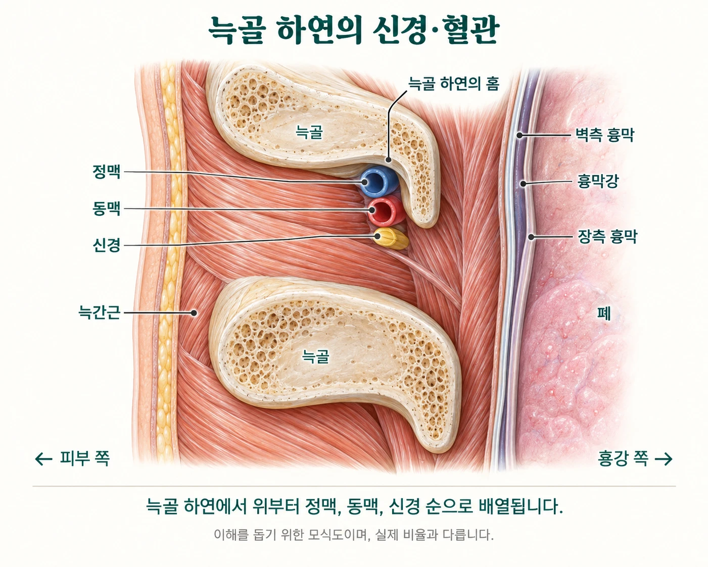
Pleura and lungs
The inner surface of the chest wall is lined by the parietal pleura, with the lung just inside. If the pleura is punctured, air enters the pleural space and the lung partially collapses. This is a pneumothorax.
Organs below the costal margin
The liver lies beneath the lower ribs on the right and the spleen on the left. These structures must be considered when working around the lower ribs.
Posterior joints
Each rib forms two joints with the spine at the back. These act as hinges, allowing the rib cage to move rather than function as a single rigid ring.