 da Re-Fit Clinic
When we inhale, the diaphragm contracts and descends while intercostal muscles and ribs expand the chest. A pressure difference draws air into the lungs. During quiet exhalation, muscles relax and recoil of the lungs and chest wall moves air out. The lungs do not inflate themselves.
Studies directly measuring postoperative lung function are discussed in our article on breathing and lung function after rib remodeling. This article explains the breathing mechanics needed to understand those results.
The lungs do not expand by themselves
It is easy to imagine that the lungs inflate first and make the chest expand. The sequence is actually the reverse.
- The diaphragm descends and the ribs rise, enlarging the chest cavity.
- Pressure inside the chest falls below outside pressure.
- That pressure difference draws air into the lungs.
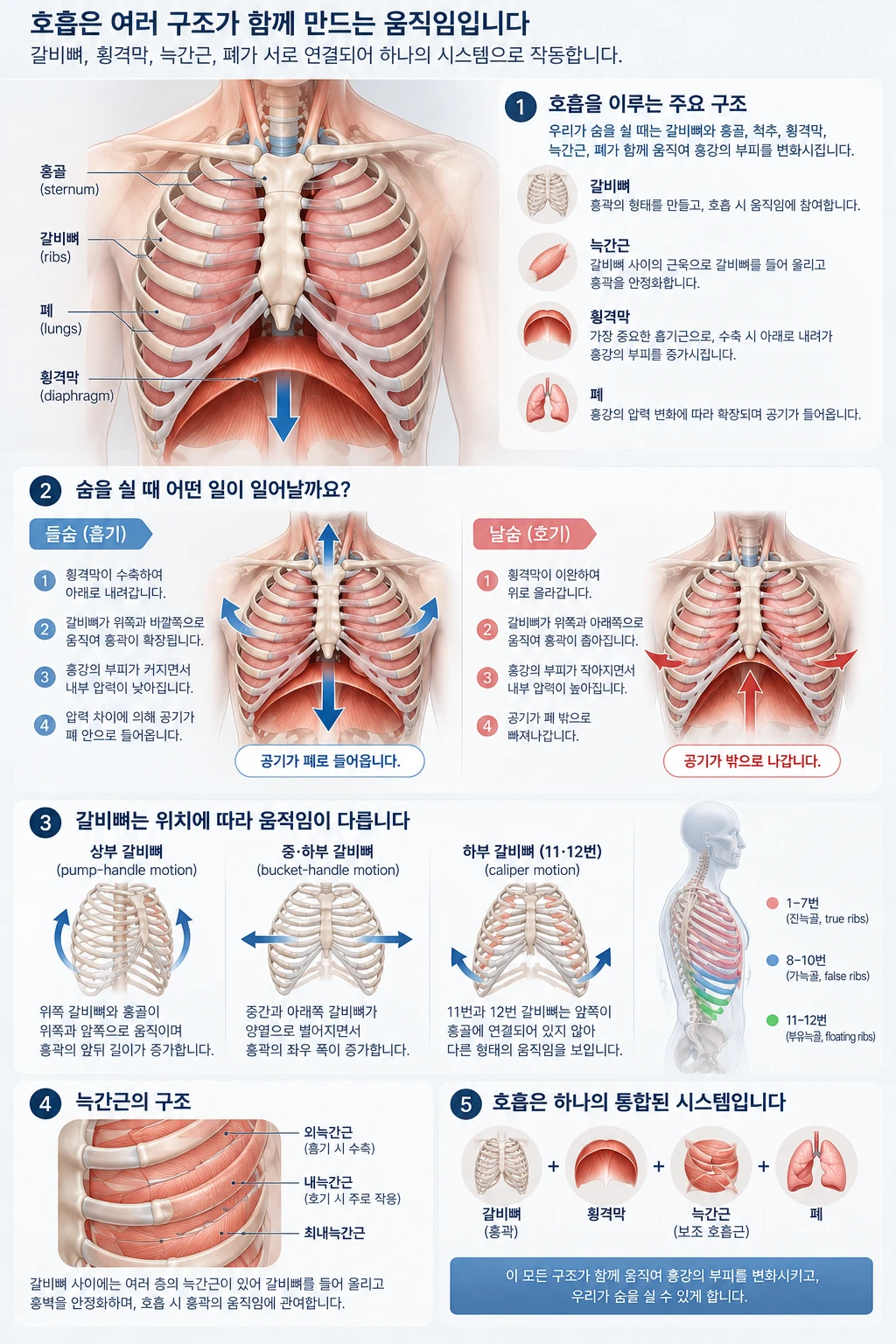
The lungs have no muscles to move themselves. They expand as the chest expands. Respiratory function cannot therefore be understood by looking only at the lungs or at a single rib.
The diaphragm is central to ordinary breathing
The diaphragm is a large dome-shaped muscle separating the chest and abdomen. On inhalation it contracts and descends, increasing the chest cavity’s vertical dimension. On exhalation it relaxes and rises, while recoil of the stretched lungs and chest wall drives air out. Quiet exhalation while seated requires very little effort.
The diaphragm’s edges attach to the inner surfaces of the lower ribs. Its contraction not only moves it down but also lifts the lower ribs upward and outward. In animal experiments, approximately 60% of its lifting force on the lower ribs came from direct muscular traction.1
Because the diaphragm and lower ribs are connected in this way, it is reasonable to ask whether lower-rib surgery affects breathing. This question has been studied; the findings are discussed again below.
Intercostal muscles lie between the ribs
The intercostal muscles move the ribs and stabilise the chest wall. Their roles differ with location and orientation. External and parasternal intercostal muscles act in inspiration, while internal intercostals act in expiration.2
In a study recording muscle activity with needle electrodes in healthy people, parasternal intercostals and scalene muscles in the neck consistently worked together even during quiet breathing. Attempting to breathe using only the diaphragm pulled the upper rib cage inward.3 Even quiet breathing is not the diaphragm’s work alone.
- The diaphragm mainly expands the chest vertically.
- The ribs and intercostal muscles help expand it front to back and side to side.
Not all ribs move in the same direction
During inhalation, ribs rise by rotating around their spinal joints. The direction varies with their position.
Part ③ of the diagram above shows three movements.
- Pump-handle motion — The front ends of the ribs and sternum rise upward and forward, increasing the chest’s front-to-back diameter. This is more prominent in the upper ribs.
- Bucket-handle motion — The sides of the ribs rise, increasing the chest’s width. This is more prominent in the middle and lower ribs.
- Caliper motion — The front ends spread sideways around the posterior joints. This describes ribs 11 and 12, whose front ends are not attached to the sternum.
In a CT study of ribs 3–7 in healthy adults, full inhalation rotated rib 3 by 11 degrees in the pump-handle direction and 13 degrees in the bucket-handle direction, and rib 7 by 7 and 10 degrees respectively.4 Dividing rib rotation into these three directions is a method used in respiratory-mechanics research.5 However, no study directly measuring how much human ribs 11 and 12 move during breathing was found.
The important point is not the names, but that the whole rib cage moves in several directions at once.
Quiet breathing differs from exercise
At rest, breathing requires little force and centres on the diaphragm. During running or breathlessness, muscles such as the sternocleidomastoid help lift the chest further, and abdominal muscles contribute to expiration.
Rib movement also changes. A CT tracking study found slightly more bucket-handle motion during quiet breathing, while pump-handle motion was approximately four times greater during full inhalation.6 The body combines muscle and rib movements according to its ventilation needs.
Does that make a single rib unimportant?
No. Each rib contributes to the chest wall, provides muscle attachments, protects organs and moves during breathing. Both of the following statements are oversimplifications:
- “There are plenty of other ribs, so losing one does not matter.”
- “Any change to a rib necessarily causes a major loss of lung function.”
Breathing is produced by the entire rib cage, diaphragm, intercostal muscles and lungs together. The effect of changing one part must be measured, not guessed.
So, is breathing preserved after rib remodeling?
The findings measured so far are as follows.
- When ribs are kept and their angles changed — In a study of 294 women aged 18–38 undergoing RIBXCAR, vital capacity and FEV1 at 6 months and 1 year did not differ significantly from preoperative measurements.7
- When ribs are removed — A study comparing 11 women who had ribs 11 and 12 removed with 11 women who had not found lower FEV1 and expiratory muscle strength in the surgical group.8
The underlying mechanics may help explain the difference. Removing a rib also removes muscle attachment sites. Angle-changing surgery retains the bone and attachment sites for the diaphragm and abdominal muscles. However, this is a mechanistic explanation; no direct comparison of the two operations is yet available.
The limitations are clear. The RIBXCAR study was conducted at one centre by the technique’s developers, with no control group and follow-up limited to one year. Participants were young women without lung disease. Breathing during exercise and respiratory muscle strength were not measured. That is why a simple reassurance is not enough. Which ribs are changed, how extensively and by which method determine the answer. → Study findings in detail
What to know about breathing after surgery
- Pain may make breathing shallow. Pain after surgery can lead to shallow breaths and incomplete lung expansion. This is why adequate pain control and deep-breathing practice matter.
- Contact the medical team immediately if sudden chest pain or breathlessness occurs. Pneumothorax is uncommon but possible and is assessed with chest imaging.
- The operation is not recommended for people with asthma or COPD. Limited respiratory reserve increases risk.
→ Risks and safety · → When surgery is not recommended
Ribs are not simply structures to remove
Ribs protect internal organs, provide muscle attachments and participate in breathing. The aim should not be to change as many ribs as possible. Understand their function, identify the structures influencing body contour, and change only what is necessary.
The waistline is not determined by a single rib either. Chest width, lower-rib direction, pelvic width, the gap between chest and pelvis, and abdominal muscles and fat all contribute. My first question is which structures create this person’s shape.
Questions for consultation
- Which ribs would be treated in my case, and to what extent?
- I have respiratory disease or a smoking history. Is surgery possible?
- How will pain be managed, and how should I practise breathing afterwards?
- If I develop breathlessness or chest pain, when and whom should I contact?
“Not how much can be changed, but how much change is necessary.”
This is the principle I consider most important in surgery involving the rib cage.
References
- Wilson TA, De Troyer A. Effects of the insertional and appositional forces of the canine diaphragm on the lower ribs. J Physiol. 2013;591(14):3539-3548. doi:10.1113/jphysiol.2013.253286
- De Troyer A, Kirkwood PA, Wilson TA. Respiratory action of the intercostal muscles. Physiol Rev. 2005;85(2):717-756. doi:10.1152/physrev.00007.2004
- De Troyer A, Estenne M. Coordination between rib cage muscles and diaphragm during quiet breathing in humans. J Appl Physiol Respir Environ Exerc Physiol. 1984;57(3):899-906. doi:10.1152/jappl.1984.57.3.899
- Wilson TA, Rehder K, Krayer S, et al. Geometry and respiratory displacement of human ribs. J Appl Physiol (1985). 1987;62(5):1872-1877. doi:10.1152/jappl.1987.62.5.1872
- Capano JG, Moritz S, Cieri RL, Reveret L, Brainerd EL. Rib Motions Don't Completely Hinge on Joint Design: Costal Joint Anatomy and Ventilatory Kinematics in a Teiid Lizard, Salvator merianae. Integr Org Biol. 2019;1(1):oby004. doi:10.1093/iob/oby004
- Luu BL, McDonald RJ, Bolsterlee B, et al. Movement of the ribs in supine humans for small and large changes in lung volume. J Appl Physiol (1985). 2021;131(1):174-183. doi:10.1152/japplphysiol.01046.2020
- Manzaneda Cipriani RM, Adrianzen GA. Respiratory Function Assessment After Waist Reduction Through Scarless Ultrasound-Guided Monocortical Fracture (RibXcar) Surgery for Body Contouring. Aesthet Surg J Open Forum. 2026;8:ojag007. doi:10.1093/asjof/ojag007
- Aburub A, Darabseh MZ, Badran R, et al. Ant-waist surgery adversely affects lung function: a cross-sectional study. Multidiscip Respir Med. 2024;19(1):984. doi:10.5826/mrm.2024.984
Rib remodeling is an elective cosmetic operation. Suitability and outcomes vary with individual anatomy and health. A decision should follow examination, testing and a thorough discussion with the medical team.
→ More on rib-cage anatomy
→ Possible complications
→ When the procedure is not recommended