 da Re-Fit Clinic
Rib remodeling requires attention not only to the outline of the ribs but also to the pleura and lungs inside them and the nerves and vessels running alongside them. Drawing on chest-tube insertion during thoracic surgery training, this article discusses chest-wall anatomy and how the scope of surgery is judged.
Opening the chest
A thoracic surgeon’s first task is to spread the ribs. To reach the heart or the lungs, that door must be opened. Opening it was where my work began.
What I learned inside was not just anatomy, but boundaries.
This far is acceptable; beyond here is not. The path of an intercostal nerve, a thin point in the pleura, an angle at which a rib must not break. I learned these boundaries, absent from a textbook, through my fingertips.
Ribs are more than bones. They are a wall around the organs. Knowing what lies inside that wall changes the way you handle it.
It was my first year of residency.
I covered both the ward and the emergency department on call. The calls never stopped. Later, I could manage dozens of cases a day quickly, but at first a single case took a long time.
That day, I had spent the day in theatre, seen patients in the emergency department and completed ward rounds. One referral from another department remained that night: a patient with fluid in the pleural space who needed a chest tube.
It was past 11 p.m.
I was close to tears because the work would not end. Still, it was my responsibility, so I went.
It was not an easy case. The patient had a high body weight and a pleural effusion, not a pneumothorax. An effusion is more difficult.
Now, inserting a chest tube takes me less than five minutes. That day it took over thirty. I kept sweating, and the patient was struggling. Even now, the memory is unsettling.
After that day, however, chest-tube insertion became easier.
For those thirty minutes, I passed through the chest wall layer by layer: skin, fat, intercostal muscle and finally the pleura. I felt my way through each layer that I would ordinarily pass through in seconds.
A textbook lists the layers of the chest wall. That day, I actually travelled through them.
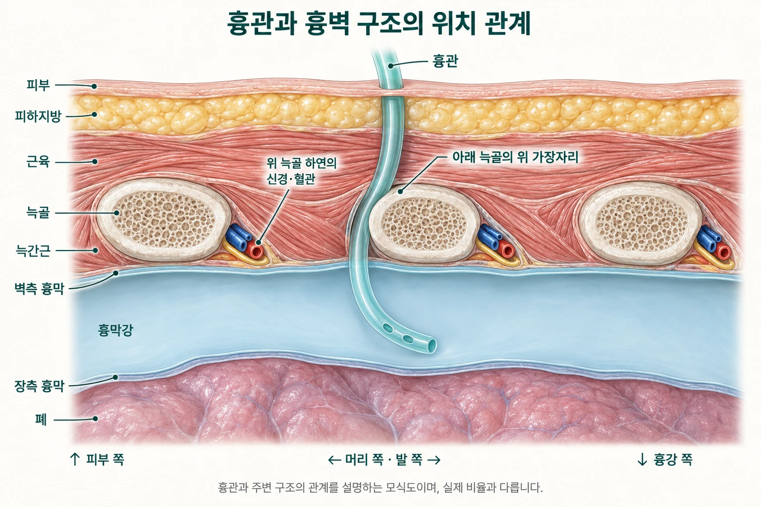
That is how boundaries are learned.
Not simply by reading, but by taking a long time, once.
Encountering the ribs again
One day, a patient asked me:
“Doctor, is it dangerous to shave down the ribs?”
I could not answer immediately. I handled ribs every day, but I did not know that particular operation.
I was surprised when I looked into it. Many people were already having it. Yet the ribs I knew felt different from the ribs described in that operation.
Looking at bone from outside is not the same as having seen it from inside.
From outside, a rib is a contour: how far it projects, differences between sides, the line toward the waist. From inside, a rib is a boundary. Beneath it are the pleura and lung, with nerves and vessels along its lower edge. The bone is the same; what you see is different.
Working with shape
A doctor who worked with function has entered the field of shape. It is not a common move.
But when it comes to ribs, I have not changed my position. I have taken one step further from where I already stood. A century of opening the chest wall, working with ribs and cartilage, and managing their complications is part of the history of thoracic surgery.
Creating a shape must not mean giving up safety. I do this work to examine whether the two can go together.
That is why I first disclose the situations in which I do not recommend the procedure: weak bones, previous rib fractures, limited respiratory reserve or fragile connective tissues.
Knowing when to stop is harder than knowing how to cut.
Knowing where to stop: that is what spreading the ribs taught me.
→ Next article · Nerves, Vessels and Cartilage: Structures to Understand Before Rib Remodeling
→ When the procedure is not recommended
→ History of Rib Surgery