 da Re-Fit Clinic
The groove along the inner lower edge of a rib carries a vein, artery and nerve. The pleura and lungs lie inside the ribs, while costal cartilage at the front contributes to chest movement. When assessing rib remodeling, the location and function of these structures matter alongside bone shape.
The tissues between the skin and ribs must be distinguished from the pleura and lungs inside. External contours alone cannot show their position and condition. Rib numbers and types are explained separately in our article on rib definition, location and structure.
Three structures run through the groove
A groove along the inner lower edge of each rib carries nerves and vessels. Within it, the vein, artery and nerve run side by side, in that order from top to bottom.
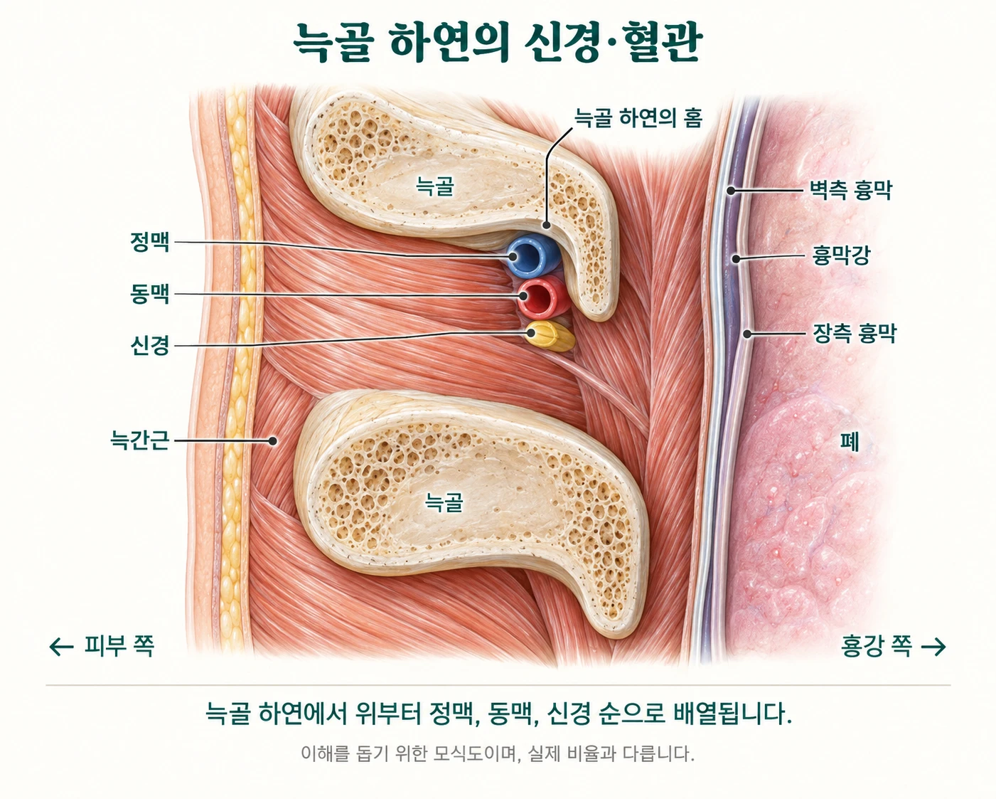
The order matters. The nerve is lowest. It sits closest to the lower edge of the rib. Force applied here affects not only the bone but also the nerve.
This is why intercostal nerve injury is a major concern when working on chest-wall shape. Pain or altered sensation can persist. Rather than hurting at one point, symptoms may form a band along a rib. Recovery can take months, and may be incomplete.
Bone is not the only tissue shaping the chest
The next lesson concerns costal cartilage. Connecting the ribs to the sternum, this tissue bends much more readily than bone and deforms under force before returning to its shape.
Take a deep breath and your chest expands. Both movement at the rib joints and the flexibility of costal cartilage contribute. This tissue helps the chest expand and contract with each breath.
For this reason, operations on chest-wall shape have focused on cartilage rather than bone over the past century. Cartilage has always guided decisions about where and how much to operate.
Its flexibility varies between people. Calcification progresses with age, so the same tissue differs between people in their twenties and forties, with considerable variation even at the same age. This is why appearance alone is not enough.
What a century has taught us
The first recorded operation on the chest wall was in 1911. A surgeon named Meyer removed two costal cartilages from a 16-year-old boy who was breathless because his chest was sunken. His symptoms remained.
Two years later, Sauerbruch removed a much larger area. Pressure on the heart was relieved and the patient returned to work, but the chest wall became so weak that the heartbeat was visible from outside.
Neither surgeon described the result as a success. Doing too little caused problems; doing too much caused problems too. The question then changed. From how much to remove, to how far it was safe to go.
Answers accumulated over a century. Ravitch established a standard in 1949; in 1998, Nuss described a way to correct chest-wall shape without removing any cartilage. Meanwhile, reconstruction after tumour removal and fixation of fractured ribs added evidence on how ribs and cartilage heal.
What does knowing the ribs mean?
Knowing the ribs is more than knowing which part projects outward.
How far can you go before chest-wall stability is compromised? Which areas affect respiratory mechanics more? What symptoms signal a problem, and what must be done? These are questions of structure and function, not just shape.
And there is one more thing. Knowing whom not to operate on.
The same procedure acts differently in people with weak bones, previous rib fractures, limited respiratory reserve or fragile tissues. Recognising this is also part of knowing the ribs.
→ More on rib-cage anatomy
→ Possible complications
→ When the procedure is not recommended