 da Re-Fit Clinic
On CT after RIBXCAR, fracture-line healing must be distinguished from changes in rib angle. A fracture line may fade as it heals, but a rib whose angle has changed does not return to its preoperative shape. No promise can be made that CT will show no trace of surgery.
“Can CT show that I have had this operation?” includes three questions: the fracture line, changes in rib shape, and recognition of a surgical history. Their evidence and limitations are considered separately below.
How does the operation treat the bone?
RIBXCAR does not remove the ribs. At the back of the lower ribs, only the outer cortex is divided. The inner cortex remains connected like a hinge as the rib is bent inward to change its angle.2 Like a fresh branch that splits on one side but stays connected on the other when bent, this is called a greenstick fracture—an incomplete fracture.
Unlike a complete fracture, the bone does not separate into two pieces. That does not mean its shape remains unchanged: changing the angle is the purpose of the operation.
- In the first study of 30 patients, rib angles measured by ultrasound changed immediately after surgery and remained smaller than before surgery at 3 months.1
- In a study of 3,805 patients from 8 countries followed for one year, the angles of ribs 10–12 decreased by an average of 10 degrees.2
“The fracture fragments do not displace” and “the shape of the bone does not change” mean different things. That distinction is the starting point here.
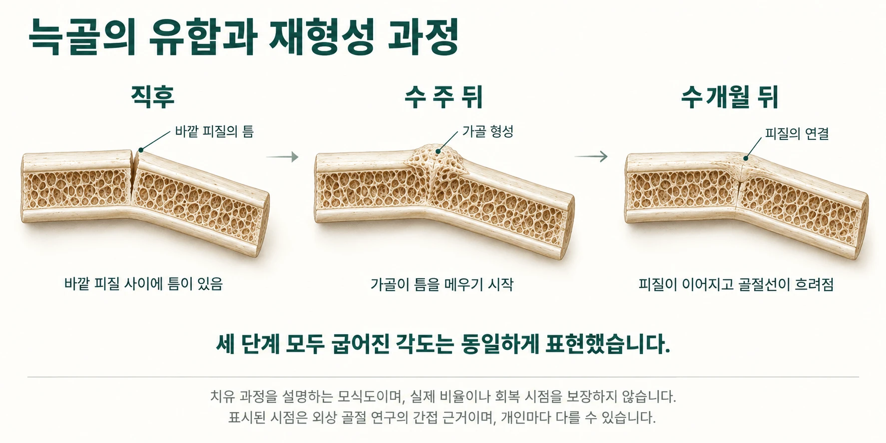
Fracture lines become less distinct with time
Studies of trauma patients show how rib fractures change on CT. These are not data from patients undergoing this operation, so the evidence is indirect.
- A study following 100 patients with traumatic rib fractures by CT over 1–6 weeks found that bony callus appeared after an average of 18 days in younger adults, 24 days in middle-aged adults and 28 days in older adults.9
- A study using CT at 3 and 6 months after metal fixation of rib fractures defined complete union as cortical continuity with the fracture line barely visible. At 3 months, only 3 of 33 fractures had complete union and 28 had partial union. All partially united fractures reached complete union by 6 months.8
- Incomplete fractures can be missed on CT even soon after injury. Of 58 rib fractures missed on initial CT readings in 130 chest-trauma patients, 56.9% were buckled rather than showing a break in cortical continuity.10
- Older fractures are detected less often. In one study, experienced radiologists’ sensitivity was 0.77 for fresh fractures, 0.61 for healing fractures and 0.55 for old fractures.11
Once the bone unites, the fracture line itself may become difficult to find, particularly with a nondisplaced incomplete fracture. However, the incomplete fractures in those studies were traumatic cortical buckling injuries, whereas this operation divides the outer cortex. They are not the same form of injury.
The changed angle remains
A fading fracture line is not the same as disappearance of every change. The angle change is an intended lasting result, and researchers use CT to assess it.
- A study of open-incision greenstick fracture showed union and inward movement of the ribs on CT at 2 months. The authors stopped corset use after confirming union on this CT.4
- A 2026 case report quantified decreased spacing between ribs on CT at 4 months.5
- A comparative study of 50 patients measured rib angles on CT before surgery and at 6 months.3
The authors of that comparative study distinguished another method that bends ribs without creating a fracture, stating that it shows no signs of union on CT because no fracture is made.3 This description assumes that signs of union can be seen with methods that create a fracture.
All of these scans were deliberately obtained to compare preoperative and postoperative appearances. The measurements were made with knowledge of what to look for and where.
Changes may be more apparent when recovery does not go as planned
If the inner cortex also breaks, producing a bicortical fracture, or the bone fails to unite and forms a pseudarthrosis, the changes can be clear.
- The 3,805-patient study reported chronic pain associated with bicortical fractures and with pseudarthrosis, which was diagnosed by CT. The authors recommended 3D CT for preoperative planning and assessment of such complications.2
- An international survey covering 2,351 patients treated by various rib-remodeling methods reported complications in 86 patients (3.7%); 15.1% of these complications involved failure of bone union.6
CT is not routinely performed after surgery
CT does not have to be obtained on a fixed postoperative schedule.
- The first study of 30 patients followed rib angles after surgery with ultrasound rather than CT.1
- The authors of a multicenter study of 131 patients did not include postoperative CT in their protocol because it could cause unnecessary radiation exposure. They stated that photographs and clinical examinations were usually sufficient, with ultrasound when further assessment was needed.12
CT is obtained when there is a reason, such as persistent pain or suspected nonunion.
What has not yet been established
- No study was found assessing whether a radiologist unaware of the surgical history could identify this operation from CT alone (PubMed search, September 2026).
- No study following CT findings beyond one year after surgery was found either. The 8 studies included in a 2025 systematic review followed patients for 3–6 months, and the authors concluded that long-term outcomes remain uncertain.7
- There are also no data describing how the corrected angle or contour at the union site changes over subsequent years in adult ribs.
The answer we can give
After union, the fracture line may become difficult to find. Changes in rib angle or at the union site may be visible on CT. Within the literature search described above, it has not been established how accurately a reader unaware of the surgery could recognise these changes.
CT is not routinely taken after surgery either. There is therefore no guarantee that surgical changes will be invisible or that a surgical history cannot be recognised.
A surgical history is not something to hide
Rib remodeling is relatively new in Korea. Many people therefore worry about later appearing to have a “surgically altered body.”
CT is used to investigate disease, not to identify who has had a particular operation. Surgical history is health information, not a trace that needs to be hidden.
If you need CT later
When having chest or abdominal CT, or seeking care for chest pain, tell your medical team when and where you had surgery. Knowing the history helps them interpret rib changes without mistaking them for trauma or another lesion.
References
- Manzaneda Cipriani RM, Duran Vega H, Cala Uribe L, et al. Waist Remodeling without Incision, with Ultrasound-guided Monocortical Fracture. Plast Reconstr Surg Glob Open. 2023;11(12):e5499. doi:10.1097/GOX.0000000000005499
- Manzaneda Cipriani RM, Duran H, Adrianzen GA, et al. A Multicenter Cohort Study of 3805 RibXcar Cases: Safety and Efficacy in Incisionless Rib Remodeling. Plast Reconstr Surg Glob Open. 2026;14(7):e7929. doi:10.1097/GOX.0000000000007929
- Manzaneda Cipriani RM, Adrianzen Nuñez GA, Medina Gutierrez JL. Waist Reduction Surgery by Rib Incurvation: A New Approach for Waist Narrowing. Plast Reconstr Surg Glob Open. 2026;14(5):e7754. doi:10.1097/GOX.0000000000007754
- Kudzaev KU, Kraiushkin IA. Waist Narrowing without Removal of Ribs. Plast Reconstr Surg Glob Open. 2021;9(7):e3680. doi:10.1097/GOX.0000000000003680
- González-Arestizábal T, Kohnenkampf R, Domínguez C. Rib remodeling as a thoracic feminization strategy in transfeminine patients: Case report. JPRAS Open. 2026;49:349-354. doi:10.1016/j.jpra.2026.02.018
- Aguilar HA, Hoyos AE, Ramirez B, et al. Global Survey on Rib Remodeling Techniques: Assessing Complications and Safety in Waistline Contouring. Plast Reconstr Surg Glob Open. 2026;14(4):e7609. doi:10.1097/GOX.0000000000007609
- Danilla S, González-Arestizábal T. Costal Surgery for Waist Improvement Safety and Efficacy: A Systematic Review of the Literature. Aesthetic Plast Surg. 2025;49(12):3433-3442. doi:10.1007/s00266-025-04815-x
- Marasco S, Quayle M, Summerhayes R, Šutalo ID, Liovic P. An assessment of outcomes with intramedullary fixation of fractured ribs. J Cardiothorac Surg. 2016;11(1):126. doi:10.1186/s13019-016-0510-3
- Hu J, Zheng ZF, Wang SH, et al. Missed rib fractures on initial chest CT in trauma patients: time patterns, clinical and forensic significance. Eur Radiol. 2021;31(4):2332-2339. doi:10.1007/s00330-020-07310-w
- Cho SH, Sung YM, Kim MS. Missed rib fractures on evaluation of initial chest CT for trauma patients: pattern analysis and diagnostic value of coronal multiplanar reconstruction images with multidetector row CT. Br J Radiol. 2012;85(1018):e845-e850. doi:10.1259/bjr/28575455
- Zhou QQ, Tang W, Wang J, et al. Automatic detection and classification of rib fractures based on patients' CT images and clinical information via convolutional neural network. Eur Radiol. 2021;31(6):3815-3825. doi:10.1007/s00330-020-07418-z
- Oñate Valdivieso C, Oñate Valdivieso D, Hoyos AE, et al. Ultrasonic- and Ultrasound-assisted Improvement of Silhouette of the Torso: Bone Structure High-definition Remodeling (Part I). Plast Reconstr Surg Glob Open. 2024;12(1):e5513. doi:10.1097/GOX.0000000000005513
Rib remodeling is an elective cosmetic operation. Suitability and outcomes vary with individual anatomy and health. A decision should follow examination, testing and a thorough discussion with the medical team.
→ What is RIBXCAR?
→ More on rib-cage anatomy
→ Possible complications
→ When the procedure is not recommended